Партнерка на США и Канаду по недвижимости, выплаты в крипто
- 30% recurring commission
- Выплаты в USDT
- Вывод каждую неделю
- Комиссия до 5 лет за каждого referral
Electroimpedance measurements of women
suffering from mastalgia
O. Trohanova *, M. Ohapkin *, A. Karpov **,
V. Cherepenin ***, A. Korzhenevsky ***
* Yaroslavl State Medical Academy, Russia
** Clinical Hospital № 9, Yaroslavl, Russia
*** Institute of Radio-Engineering and Electronics of Russian
Academy of Sciences, Moscow, Russia
e-mail: sim-tech@*****
Abstract. We examined 320 women (121 – normal state, 113 – mastitis, 86-mastalgia). To visualize the tissue of a mammary gland we used a 256 channel electro-impedance mammograph “МЕИК” (Russia). The age of patients was taken into account. An impedance image of the mammary gland in case of mastalgia is shown within a grey scale with smooth transitions from dark to light sections i. e. from low to higher conductivity without foci and where a zone of lactic sinuses is clearly singled out. In corresponding age groups women with mastalgia have the same conductivity in the left and right mammary glands while lying or standing. The values are the same in the first and second phase of a menstrual cycle. You can trace a certain increase of conductivity with age. During investigations while comparing conductivity in all age groups with chronic cystic mastitis in both positions with the one in case of mastalgia the latter turned out higher. It proves that mastalgia and chronic cystic mastitis are two different clinical conditions of the mammary gland. The results of our investigations allow us to say that electro-impedance mammography can be used as a method for differential diagnosing the normal state and different clinical conditions of the mammary gland.
Index Terms – Breast tissue diagnostics, electrical impedance tomography.
1. Introduction
Mastalgia, (mastodynia, Cooper’s disease) is a pain in the breast gland. Women, suffering from mastodynia experience pain and feeling discomfort, which become more intensive before menstruation. It is assumed, that the reason for mastalgia is a cyclic hardening of the mammary glands, caused by venous congestion and edematous stroma before menstruation, due to a change in the hormonal background; during this period the mammary gland increases in volume by more than 15 %. No reliable data have been discovered yet on the cause of mastalgia: it is not known for certain whether it is a specific state of the mammary gland or is caused by the hormonal background or by some pathology.
In the work under consideration we decided to examine the state of the mammary glands during mastalgia, using the method of bioimpedance mammography, and give a comparative estimation of the obtained parameters with normal states as well as cases of fibrous - cystic mastopathy. We had already proved, that electroimpedance mammography make it possible to receive the image and the digital data (electorconductivity, histogram) of the mammary glands’ state during various physiological periods a woman’s life (1st phase, 2nd phase of the menstrual cycle, pregnancy, lactation, postmenopause), as well as allows to diagnose fibrous - cystic mastopathy.
2. Materials and methods
We carried out examination of 320 women that comprised 3 clinical groups:
- 1st group - 121 woman aged from 17 to 76 years without pathological changes in mammary glands. The group was divided according to the age subgroups: up to 30 years - 45 persons, years - 32 persons, 41-50 years - 20 person, after 50 years - 24 persons;
- 2nd group - 86 women aged from 17 up to 50 years diagnosed as suffering from "Mastalgia" (Mastodynia) who according to the results of radiological mammography did not have pathological changes of mammary glands, but according to the results of ultrasonic exmination had certain expansion the ducts. The group was divided into the following age subgroups: up to 30 years - 46 persons, 31-40 years - 24 persons, 41-50 years - 16 persons;
- 3rd group - 113 women aged from 21 up to 64 years with the diagnosis of “Fibrous - cystic mastopathy”, established during ultrasonic and radiological mammography. The group also has been divided into the following age subgroups: up to 30 years - 12 persons, 31-40 years - 44 persons, 41-50 years - 36 person, after 50 years - 21 persons.
The 256-electrode electroimpedance mammograph, developed by the Institute of Radio - Engineering and Electronics of the Russian Academy of Science, with frequency of 50 KHz was used for visualization of the mammary gland puter scanning of mammary glands was performed at the step of every 8 cm and the depth up to 6 cm (seven planes of scanning) with the patient in recumbent or standing position. Preliminary to the examination, the mammary glands had been moistened. Duration of one patient’s examination amounted to 10-15 minutes.
The analysis of the received EIM comprised: a visual estimation of the image; acquisition of the generalized parameters of electroconductivity and its variability (average value, standard deviation); methods of variational statistics (Student’s criterion), an estimation of graphic distribution of electroconductivity and comparison of the received data in the three clinical groups.
3. Result
Electroimpedance measurements were carried out during the 1st stage of the menstrual cycle (5-10 days), during 2nd phase of the menstrual cycle days).
3.1. Electroimpedance image of mammary glands of women suffering from mastalgia.
3.1.1. Qualitative estimation of the electroimpedance image.
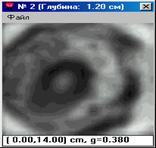
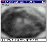
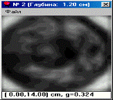
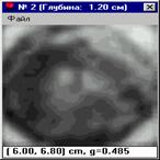
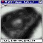
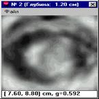
The electroimpedance image of the mammary gland with mastalgia is represented by gradation of the grey scale with smooth transitions from dark (hyperimpedance) to light (hyporimpedance) areas, i. e. from low to high electroconductivity. Absence of focus formation, i. e. prominent areas with sharp edges is significant. The mammary glands images of practically all menstruating women during the whole menstrual cycle (during the first as well as the second phases) show very distinctly the postmammilla area or the so-called zone of mammary sinus in the form of a rounded hypoimpedance or hyperimpedance area with the indistinct contours gradually blending into surrounding tissues. More often this zone is prominent at the 1st and the 2nd levels of scanning, i. e. at the depth of 0,4 - 1,2 cm and less often at the 3d level of scanning, i. e. at the depth of 2 cm, but beginning from the 4th to the 7th levels of scanning the zone of mammary sinus doesn’t stand out (the depth of scanning 2,8 - 5,2 cm). Having analyzed all images we were enable to discover any visual difference between the left and the right mammary glands, with the women neither in a recumbent nor in standing position. Therefore, figure 1 represents electroimpedance images of mammary glands during mastalgia, based only on the women’s age and a phase of the menstrual cycle.
The electroimpedance images during various phases of menstrual cycle practically do not differ from one another; the amount the grey scale dark tones - the hyperimpedance areas, characterizing low electroconductivity of glandular tissues, decreases with age, and the amount of the light tones in the grey scale, characterizing high electroconductivity of connecting and hyaline tissues, increases. It proves presence of involutive changes in the mammary gland tissues at mastalgia.
3.1.2. Quantitative estimation of the electroimpedance image at mastalgia.
In order to estimate electroconductivity of mammary gland tissues we used data from 2nd level of scanning (depth of scanning about 1,2-2,0 cm). Electroconductivity of tissues is expressed in conventional units.
During detailed examination we found no differences in electroconductivity between the left and the right mammary glands of women suffering from mastalgia in corresponding age groups either in the 1st or in 2nd stage. The difference in all cases is statistically not valid (p> 0,05). No statistic difference of the mammary glands electroconductivity was observed either in recumbent or in standing position as well. (p> 0,05). Therefore, for further work and comparative estimation, we used parameters of average electroconductivity in both phases of menstrual cycle of women in a recumbent position. The received results are submitted in table 1.
Table 1. Average electroconductivity of mammary glands in different age periods of women with mastalgia (recumbent position) (М m).
Age groups | Up to 30 | 31-40 | 41-50 |
Average an electoconductivity in the 1st and 2nd phases of the cycle | 0,39±0,11 | 0,41±0,15 | 0,5±0,08 |
The increase of electroconductivity parameters with aging is apparent. There is a valid difference in the average electroconductivity of mammary glands during mastalgia in different age groups “up to 30” – “41-50” and “31-40” – “41-50” (t1-3 = 5,05, p <0,01; t2-3 = 3,02, p <0,01;). In the groups “up to 30” – “31- 40” - the difference is not statistically valid (t1-2 = 0,9, p> 0,05).
3.2. Comparative estimation of electroimpedance images made during mastalgia, in normal cases and fibrous - cystic mastopathy (FCM).
3.2.1. Qualitative estimation of electroimpedance images made during mastalgia, in normal cases and fibrous - cystic mastopathy (FCM).
If electroimpedance images in norms are compared with those made during mastalgia and mastopathy in all age groups, one can observe presence of darker tones of the grey scale and hyperimpedance areas in the electroimpedance image that belong to women with fibrous - cystic mastopathy.
3.2.2. Quantitative estimation of electroimpedance images made during mastalgia, in normal cases and fibrous - cystic mastopathy (FCM).
In our earlier publications as well as reports we performed estimation of an electroimpedance image in norms and at fibrous - cystic mastopathy. We were able to prove absence of statistically valid difference between the left and right mammary glands, taken with women in recumbent and standing positions during various phases of menstrual cycle in corresponding age groups. Therefore, in order to carry out comparison of electroconductivity at mastalgia with norms and fibrous - cystic mastopathy we used earlier obtained average indices of electroconductivity.
The results are shown in table 2.
Table 2. Average electroconductivity of mammary glands during different age periods of women in normal cases, with mastalgia, and fibrous - cystic mastopathy in recumbent position (М m).
Age groups | Norm | Mastalgia | FCM |
Up to the age of 30 | 0,34±0,09 | 0,3±90,11 | 0,27±0,09 |
31 – 40 years old | 0,4±60,1 | 0,41±0,15 | 0,36±0,12 |
41 – 50 years old | 0,46±0,1 | 0,5±0,08 | 0,4±40,1 |
It is evident from the table, that the parameters of the average electroconductivity of mammal glands of women in a recumbent position, suffering from mastalgia, in the age groups of “up to 30 years ” and “41-50 years” are higher, than in normal cases, which is statistically valid (“up to 30 years” - t1-2=3,35, p <0,01; “41-50 years ” - t1-2=1,9, p <0,05); and in the age group of “31-40 years” it is statistically valid that electorconductivity is lower at mastalgia (t1-2=2,11, p <0,05). Parameters of the average electroconductivity in a recumbent position at mastalgia in all age groups are higher than parameters of the average electroconductivity in case of fibrous - cystic mastopathy. The conclusion is also statistically valid (“up to 30 years ” - t2-3=4,92, p <0,01; “31-40 years ” - t2-3=2,12, p <0,05; “ 41-50 years ” - t2-3=2,92, p <0,01).
4. Discussion
Thus:
1. Mammary glands electroconductivity in case of mastalgia depends neither on the scanning side nor on the patient’s position during examination.
2. No statistically significant distinctions in electroconductivity of mammary glands were revealed during examination of women with mastalgia at different stages of the menstrual cycle.
3. The comparison of electroimpedance images of the mammary glands in norm, with mastalgia and fibrous - cystic mastopathy of all age groups revealed presence of darker tones of the grey scale and hyperimpedance areas in the electroimpedance images of women with fibrous - cystic mastopathy, irrespective of the stage of the menstrual cycle. It means that the changes more often have a defuse character, involving all anatomic structures of the mammary gland, without focal symptomatology. This statement is confirmed by the graphic characteristic of the electroconductivity distribution in the electroimpedance image of normal mammary glands, glands with mastalgia as well as fibrous - cystic mastopathy where unimodal distribution of electroconductivity prevails.
5. Having analyzed the electroconductivity during various age periods, we discovered the following tendency: the electorconductivity of the mammary glands increases in case of healthy women as well as women with mastalgia and fibrous - cystic mastopathy, this is statistically valid and reflects connection of pathological changes of tissues at different stages of evolution. But comparison of the mammary glands electroconductivity in different clinical groups revealed that characteristics of electroconductivity are statistically significantly lower in cases of fibrous - cystic mastopathy, and electroconductivity at mastalgia occupies an intermediate position between norms and fibrous - cystic mastopathy. It once again proves that mastalgia and fibrous - cystic mastopathy are two different states of the mammary gland resulting from a dysfunction in the system “ovaries - mammary gland”, which cannot be classified as separate diseases.
6. The conducted research makes it possible to use electroimpedance mammography as a method of differential diagnostics of normal mammary gland as well as its various conditions.
5. The literature
1. Gubler E. Calculation methods of analysis and recognition of pathological processes, 1978.
2. A. Karpov, V. Cherepenin, V. Korjenevsky and A. Mazaletskaya Qualitative estimation of electro-impedance tomogram 1st EPSRC Engineering Network meeting. london, 1999.
3. A. Korjenevsky, V. Cherepenin and V. Kornienko Electric Mammograph with 3D visualization. 1st EPSRC Engineering Network meeting. London, 1999.
4. Nyboer J. Bagno S, Nims LF. The electrical impedance plethysmograph an electrical volume recorder. Washington, DC: National Research Council, Committee on Aviation, 1943. (Repon no 149.)
5. Pethig R. Dielectric and electronic properties of biological materials. New York: John Wiley and Sons, 1979.
6. Stanton A. Glantz Primer of Biostatistics, 1999.
7. O. Trokhanova, A. Karpov, M. Ochapkin, V. Cherepenin, A. Korjenevsky, V. Kornienko, Y. Kultyasov, Electro-impedance mammography testing at some physiological woman's periods. XI international conference on electrical bio-impedance. Oslo, Norway, 2001.
8. O. Trokhanova, A. Karpov, M. Ochapkin, V. Cherepenin, A. Korjenevsky, V. Kornienko. Electro-impedance mammography testing at some physiological woman’s periods. 3rd EPSRC Engineering Network meeting. London, UK, 2001.
2nd phase. Up to 30 years 2nd phase. 31-40 years 2nd phase. 41-50 years

2nd phase. Up to 30 years 2nd phase. 31-40 years 2nd phase. 41-50 years
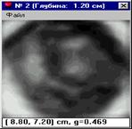
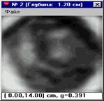
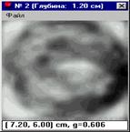
Figure 1. Electroimpedance image of mammary glands of women from different age groups, suffering from mastalgia, during various phases menstrual cycle.
Norm Mastalgia FCM