
Партнерка на США и Канаду по недвижимости, выплаты в крипто
- 30% recurring commission
- Выплаты в USDT
- Вывод каждую неделю
- Комиссия до 5 лет за каждого referral
THE STUDY OF CHROMOSOME DAMAGES IN THE HUMAN CELLS IRRADIATED BY THE THERAPEUTIC PROTON BEAM
Zaytceva E. M., Govorun R. D.
Joint Institute for Nuclear Research, Dubna, Russia
The therapeutic proton beam was based on the phasotron in LNP and has been used for a long period for radiation therapy (Mytsin G. V. et al, 2002). Peculiarities of the proton energy deposition and the Bragg peak at the end of the particles path allow us to form an optimal dose in the tumour tissues.
The main object of investigation was the study of peculiarities of chromosomal aberrations’ formation and the estimation of efficiency of the therapeutic proton beam with initial energy of 170 MeV and in the Bragg peak region.
Materials and Methods
The chromosome aberrations in the human cells (on a model of blood lymphocytes) were studied after irradiation by 170 MeV proton beam prepared for therapy of patients on the entrance and at the Bragg peak region, that corresponds to the irradiation of normal tissues along the beam path and tumour tissues directly. The energy of the Bragg peak protons is the spectrum from 0 to 30 MeV. The spectrum of LET stretches up to ~ 100 keV/mkm. The unstable chromosome aberrations were analysed by the metaphase method.
Results and Discussion
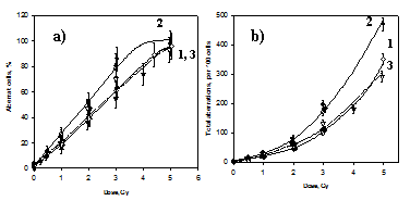 |
Protons and γ-rays induce the chromosome aberrations in the lymphocytes. As it has been seen (Fig.1a), the frequency of aberrant cells increases linearly from dose up to ~ 4,5 Gy of 170 MeV protons and γ-rays and to 3,5 Gy of the Bragg peak protons (to 90 % of the aberrant cells). Total number of chromosome aberrations increases exponentially from the doses (Fig.1b).
Figure 1. Dose-dependences of aberrant cells frequency (a) and total number of chromosome aberrations (b) after irradiation by 170 MeV protons (1), the Bregg peak protons (2) and γ-rays (3)
High efficiency of the Bragg peak protons has been shown. RBE values were ~ 1,25 ( at the dose interval of 1-4 Gy) as it was estimated at these two tests, while the protons with energy of 170 MeV did not differ from the γ-radiation. Our data are in a good agreement with the results obtained by the authors (Vitanova A. et al, 2002). Also the fraction of cells with numerous aberrations (3 and more) exceeded ~3 times (27% and 10%). The high level of exchange chromosome aberrations (up to 75%) with a predomination of dicentrics (~50 %) was revealed in the analysis of different aberrations.
As the proton beam dose to a tumour is formed from several directions (up to 7), the damage of cells in surrounding tissues along protons path will decrease. So after the dose of 3 Gy about 80% of the tumour cells obtain damages, but in surrounding healthy tissues it will not exceed 10% (Fig.1a).
Conclusion
Our investingation confirms a high efficiency of proton beams for use in radiation therapy.
References
1 MYTSIN G. V. et al. 2002. Dubna centre of hadron therapy, status and perspectives.// 3-rd Rus. Science forum “Radiation diagnostics and radiation therapy in a clinic of XXI c.”. P. 109 (Rus).
2 VITANOVA A. et al. 2002. The study of Relative Biological Efficiency of clinical phasotron proton beam of LNP JINR // Preprint JINR. P. P. 1-7.(Rus)
