
Партнерка на США и Канаду по недвижимости, выплаты в крипто
- 30% recurring commission
- Выплаты в USDT
- Вывод каждую неделю
- Комиссия до 5 лет за каждого referral
24
Трехмерная структура белков
The Protein Data Bank contains more than 2000 NMR structure of proteins and nucleic acids.
Molecular mass distribution of the NMR structure deposited in the Brookhaven national Bank, as of December 1997. The number of structures deposited is plotted versus the molecular mass (Guntert, 1998). The figure clearly shows that the most NMR structures are in the 1 to 25 kDa range, with maximum at 7 to 9 kDa and only a few structures with molecular masses above 25 kDa. The scarcity of NMR structures above 25 kDa manifests the fact that conventional NMR spectroscopy in solution can not readily be used with structures that have so large molecular masses.
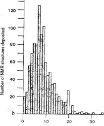
Молекулярная масса белка в кDа
The foundations of solution NMR structural studies are:
high-quality NMR spectra recorded with good sensitivity and spectral resolution.
However, with increasing molecular mass, these basic requirements are difficult to achieve. Limiting factors are:
low sensitivity and line broadening and extensive signal overlap due to the high complexity of the spectra.
Small proteins (6-10 kDa)
Wuthrich, (2001) The way to NMR structures of proteins, Natur. Struct. Biol., 8, 923-926, 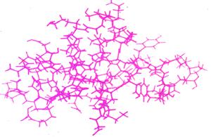
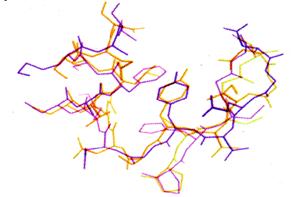
The first protein structure determined by NMR. a. All heavy-atom presentation of the NMR structure of the proteinase inhibitor IIA from bull seminal plasma (BUSI IIA). b. Superposition of the core region of residues 23-42 in the NMR structure of BUSI IIA (green) with the corresponding polypeptide segment in the X-ray crystal structure of the homologous porcine pancreatic secretory trypsin inhibitor (PSTI) (blue)
