

Партнерка на США и Канаду по недвижимости, выплаты в крипто
- 30% recurring commission
- Выплаты в USDT
- Вывод каждую неделю
- Комиссия до 5 лет за каждого referral
UDK 616.441-018-097:616.149-008.341.1
T. M. Dosayev, S. N. Romaniuk, S. T. Omarova, B. Zh. Salimgereyeva, A. S. Semyenov
S. Asfendiyarov Kazakh National Medical University, Almaty, Kazakhstan
Morphologic and Immunohistochemical Description of the spleen of the Patients with Portal Hypertension Syndrome
Abstract. Immunohistochemical analysis of T-cellular compartments of the spleen has been carried out on autopsy material of the patients died with clinical diagnosis of liver cirrhosis.
The results of investigation have shown, that structural functional changes, manifesting in a significant decrease of volumetric density of the white pulp and disturbance of the processes of T-lymphocytes differentiation, promoting T-cellular immunodeficit, occur in the spleen of the patients with the syndrome of portal hypertension.
Key words: immunohistochemistry, spleen, portal hypertension, T-lymphocytes.
Introduction
According to WHO data approximately 300 thousand men all over the world develop viral hepatitis B annually. Every year more than 200 million of people are considered to be HIV carriers from whom about 2 million die because of hepatitis B infection, 650000 because of cirrhosis and liver cancer as a result of chronic infection.
Liver cirrhosis also develops in 10-20 % people abusing alcohol.
At present considerable progress has been achieved in investigation of the immune system organs and, in particular, the spleen (1, 2, 3, 4, 5). Nevertheless, many intricate questions still exist round morphology and function of the man’s spleen. It is explained by a number of reasons. The spleen is extremely sensitive to autolysis that impedes understanding autopsy material obtained after death. Great problem in interpretation and definition of the spleen structure in health is also connected with the fact that it is not always known what diseases, affecting immunogenesis function, a man underwent during his life and what antiviral vaccination he got in his childhood. Another problem consists of a quick spleen constriction after death bounded up with a sharp pressure falling in the splenic vein and in the portal system as a whole, and morphologic description of the sections of such a spleen does not correspond to its structure in the living organism. In this connection further investigations of the spleen of man in health and pathology are necessary.
Aim of the research – to study morphology and functional specific characteristics of immunocompetent cells of the spleen in patients with portal hypertension syndrome.
Material and methods
Investigation was carried out on autopsy material of 24 adult patients of the 1 st and 2 nd periods of mature age, died with clinical diagnosis of liver cirrhosis in the stage of decompensation, according to the generally accepted scheme of ontogenesis periodicity of a man.
Two cases of splenectomy of persons who perished as a result of trauma and without signs of hypersplenism and blood diseases in anamnesis were taken as a control.
Morphologic analysis of the spleen parenchlyma was conducted on preparations stained with hematoxylin and eozine, as well as azure-2 and eozine. Volumetric density of the white pulp was determined by means of standard ocular net LOMU( Leningrad Optical Mechanical Union ).
Serial paraffin spleen sections of 5 mcm thickness fixed in formalin were stained immunohistochemically with antibodies on lymphocytes markers with receptors CD1a+, CD+, CD4+, CD8+, CD10+, CD20+, CD23+, as well as on myeloperoxidase to reveal neutrophiles and eozinofiles.
Decamouflage was carried out in buffer solution pH9,0 in apparatus PT LINK DAKO for 20 minutes at temperature +97C. Arrangement of the reaction was conducted in Autostainer Link 48 DAKO using visualization system En Vision Flex+Mouse (Link). Tincturing was in En Vision FLEX HEMATOXLIN.
Dehydration was in three ethanol changes and enlightenment in O-xylene. Then paraffin spleen sections were put into tegumental medium MOUNT-QUICK.
Results of the research
Local indurations of the reticular fibers without vessels, formed after postmortem collapse of the spleen in connection with sharp falling of the venous blood pressure in the portal system were observed in the red pulp along the spleen parenchyma of man in the control. T-dependent and B-dependent zones were clearly defined in the lymphoid nodes, located eccentrically from the central artery (fig.1).
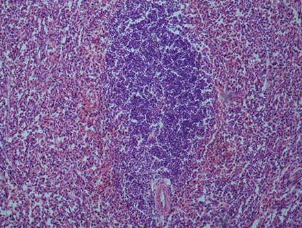
Fig. 1. Control. Lymphoid node of the spleen of a man. Hematoxyline and eosin. Objective 20, ocular 10.
T-lymphocytes (CD3+) were situated round arterioles and represented themselves spaces of irregular form occupied with small polymorphous lymphocytes, the majority of which were stained immunohistochemically with cluster CD4+ (T helpers) antibodies. Hardly defined perifollicular zone was also visually observed round T-lymphocytes accumulations.
Lymphoid nodes bordered on with T-lymphocytes of periarterial lymphoid sheath, dividing with them common perifollicular zone. Branches of arterioles passed not only through T-dependent zones, but through B-dependent zones of the lymphoid nodes, including germinal centers. Sharp decrease of volumetric density of the white pulp -11,7±0,4 (in the control 21±0,4) p<0,05 and sharp enlargement of the connective tissue, particularly in the places of the lymphoid nodes localization, have been revealed in the spleen of the patients with liver cirrhosis.
Single lymphoid nodes, met in parenchyma in the red pulp, were not large in size and represented only accumulations of the lymphocytes without clear separation into T and B – compartments, as they consisted practically of only T-lymphocytes alone (fig. 2).
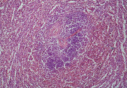
Fig. 2. Portal hypertension. Lymphoid node of the man’s spleen. Hematoxyline and eosin. Objective 20, ocular 10.
Such lymphoid nodes didn’t have clearly marked marginal zone, germinate centre and crown, clear definite border between nodes and red pulp was completely absent as well. Periarterial lymphoid sheath, typical for the spleen in health, being one of the main components of the white pulp, was completely absent round pulpal vessels.
Sharp deficit of the general T-lymphocytes pool (mature functionally active CD+3 cells) against a background of increased quantity of cytotoxic cells T-killers (CD+), closely located along venous sinuses forming crisscross plexuses, the so-called “crisscross vessels” and in the walls of the pulpal veins, was detected in the spleen parenchema at histochemical analysis of immunocompetent cells (fig. 3).
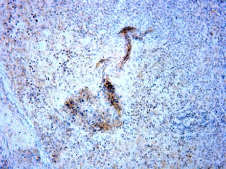
Fig. 3. Portal hypertension. CD+8 lymphocytes along venous sinuses “crisscross vessels”. Objective 20, ocular 10.
In that way, the results of the research have shown that structural-functional changes, manifesting in a significant decrease of volumetric density of the white pulp and disturbance of the processes of T-lymphocytes differentiation, promoting the development of T-cellular immunodeficit, occur in the spleen of the patients with the syndrome of portal hypertension.
Prospects
Investigations of the immune system organs in case of portal hypertension will enable to detect mechanisms of immunogenesis disturbances for the subsequent immunocorrection in patients with the syndrome of portal hypertension.
Literature
1.Mebius RE, Kraal G. Structure and function of the spleen//Nat Rev Immunol.-2005.-№5.-606-616
2.Cesta M. F. Normal Structure, Function, and Histology of the Spleen//Toxicologic Pathology.-2006.-vol.34.-5.-455-465
3.Randall K. J., Pearse G. A dual-label technique for the immunohistochemical demonstration of T-lymphocyte subsets in formalin-fixed, paraffin-embedded rat lymphoid tissue// Toxicologic Pathology.-2008.-vol.36.-795-804
4.Klatt E. Atlas of Patology. Florida State University (пер. на русский. «Логосфера».-2010.-С.-1-531)
5., , Дьяконов характеристика селезенки человека (иммуногистохимическое исследование)//
Морфология (С-Петербург).-2013.-3.-27-31
МОРФОЛОГИЧЕСКАЯ И ИММУНОГИСТОХИМИЧЕСКАЯ ХАРАКТЕРИСТИКА СЕЛЕЗЕНКИ БОЛЬНЫХ С СИНДРОМОМ ПОРТАЛЬНОЙ ГИПЕРТЕНЗИИ
, ,
Резюме. На утопсийном материале от больных, умерших с клиническим диагнозом – цирроз печени в стадии декомпенсации, проведен иммуногистохимический анализ Т-клеточных компартменов селезенки.
Результаты исследования показали, что у больных с синдромом портальной гипертензии в селезенке происходят структурно-функциональные перестройки, выражающиеся в значительном уменьшении объемной плотности белой пульпы и нарушении процессов дифференцировки Т-лимфоцитов, способствующие развитию Т-клеточного иммунодефицита.
Ключевые слова: иммуногистохимия, селезенка, портальная гипертензия, Т-лимфоциты
Казахский Национальный медицинский университет им. С. Асфендиярова
г. Алматы, Казахстан
МОРФОЛОГІЧНА ТА ІМУНОГІСТОХІМІЧНА ХАРАКТЕРИСТИКА СЕЛЕЗІНКИ ХВОРИХ З СИНДРОМОМ ПОРТАЛЬНОЇ ГИПЕРТЕНЗІЇ
єв, , імгереєва,
Резюме. На аутопсійному матеріалі хворих, які померли з клінічним діагнозом – цироз печінки у стадії декомпенсації, було проведено імуногістохімічний аналіз Т-клітковинних компартментів селезінки.
Результати дослідження показали, що у хворих з синдромом портальної гіпертензії в селезінці відбуваються структурно-функціональні перебудови які проявляються у значному зменшенні об’ємної щільності білої пульпи та порушенні процесів диференціювання Т-лімфоцитів, що сприяють розвитку Т-клітковинного імунодефіціта.
Ключові слова: імуногістохімія, селезінка, портальна гіпертензія, Т-лімфоцити.
Казахський Національний медичний університет ім. С. Асфендіярова, місто Алма-Ати, Казахстан
