
Партнерка на США и Канаду по недвижимости, выплаты в крипто
- 30% recurring commission
- Выплаты в USDT
- Вывод каждую неделю
- Комиссия до 5 лет за каждого referral
Зачетная работа по 1 блоку 2 семестра.
RISA_ECOLI, рекомендуемое название - Альфа цепочка рибофлавинсинтазы (Riboflavin synthase alpha chain) это белок бактерии Escherichia coli (кишечная палочка), находящийся в цитоплазме и участвующий в биосинтезе рибофлавина (витамин B2).
На рисунке представлена третичная структура белка (по записи 1PKV банка PDB):
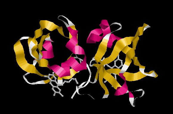
Красным покрашены альфа-спирали, желтым – бета-тяжи. Также изображены молекулы рибофлавина, выступающие в роли лигандов.
Рибофлавинсинтаза - это бифункциональный ферментный комплекс, катализирующий образование рибофлавина из 5-амино-6-(1'-D)-рибитил-амино-2,4(1H,3H)-пиримидина и L-3,4-дигидрохи-2-бутанон-4-фосфата с помощью 6,7-диметил-8-лумазина. Этот комплекс состоит из 3-х альфа-субъединиц и 60-ти бета-субъединиц. Альфа-субъединица (данный белок) катализирует дисмутацию (диспропорционирование) 6,7-диметил-8-лумазина в рибофлавин и 5-амино-6-(1'-D)-рибитил-амино-2,4(1H,3H)-пиримидин.
Данный белок является ферментом и относится к классу трансфераз. EC=2.5.1.9 по международной классификации ферментов.
Катализируемая реакция:
2 (6,7-диметил-8-(1-D-рибитил)лумазин) = рибофлавин + 4-(1-D-рибитиламино)-5-амино-2,6-дигидроксипиримидин.
Аминокислотная последовательность белка в fasta-формате:
>RISA_ECOLI Riboflavin synthase alpha chain
MFTGIVQGTAKLVSIDEKPNFRTHVVELPDHMLDGLETGASVAHNGCCLTVTEINGNHVSFDLMKETLRITNLGDLKVGDWVNVERAAKFSDEIGGHLMSGHIMTTAEVAKILTSENNRQIWFKVQDSQLMKYILYKGFIGIDGISLTVGEVTPTRFCVHLIPETLERTTLGKKKLGARVNIEIDPQTQAVVDTVERVLAARENAMNQPGTEA
Длина последовательности 213 аминокислотных остатка. Молекулярная масса 23445 Дальтон. Содержит 2 лумазин-свзязывающих повтора (с 1 по 97 и с 98 по 195 а. о.).
Ссылки на записи про белок в различных банках данных.
PDB: 1HZE, 1I18, 1I8D, 1PKV.
UniProt: P0AFU8, P0AFU9.
Ссылки на статьи, посвященные белку:
Show 'large scale' references »[1]"Cloning, sequencing, mapping and hyperexpression of the ribC gene coding for riboflavin synthase of Escherichia coli."
Eberhardt S. M.R., Richter G., Gimbel W., Werner T., Bacher A.
Eur. J. Biochem. 242:712-719(1996) [PubMed: 9022701]
Аннотация:
The gene coding for riboflavin synthase of Escherichia coli has been cloned by marker rescue on a 6-kb fragment that has been sequenced. The riboflavin synthase gene is identical to the ribC locus and codes for a protein of 213 amino acids with a mass of 23.4 kDa. It was mapped to a position at 37.5 min on the physical map of the E. coli chromosome. The 3' end of the ribC gene is directly adjacent to the cfa gene, which codes for cyclopropane-fatty-acid synthase. This gene is followed by two open reading frames designated ydhC and ydhB, which are predicted to code for putative proteins with 403 amino acids and 310 amino acids, respectively. The gene ydhC is similar to genes coding for resistance against various antibiotics (cmlA, bcr) and probably codes for a transmembrane protein. The protein specified by ydhB shows sequence similarity to a large family of DNA-binding proteins and probably represents a helix-turn-helix protein. The ydhB gene is directly adjacent to the regulatory gene purR. A 288-bp segment of the cfa gene has earlier been mapped incorrectly to a position adjacent to greA at 67 min. The ribC gene was hyperexpressed in recombinant E. coli strains to a level of about 30% of cellular protein. The protein was purified to homogeneity by chromatography. The specific activity was 26000 nmol. mg-1.h-1. The protein sediments at a velocity of S20 = 3.8 S. Sedimentation-equilibrium centrifugation indicated a molecular mass of 70 kDa, consistent with a trimer structure. The primary structure of riboflavin synthase is characterized by internal sequence similarity (25 identical amino acids in the C-terminal and N-terminal parts suggesting two structurally similar folding domains.
[2]"Analysis of the boundaries of Salmonella pathogenicity island 2 and the corresponding chromosomal region of Escherichia coli K-12."
Hensel M., Shea J. E., Baeumler A. J., Gleeson C., Blattner F. R., Holden D. W.
J. Bacteriol. 179:1105-1111(1997) [PubMed: 9023191]
Аннотация:
We recently identified a pathogenicity island (SPI2) located at 30.7 centisomes on the Salmonella typhimurium chromosome. SPI2 contains genes encoding a type III secretion system whose function is distinct from that of the type III secretion system encoded by a pathogenicity island (SPI1) at 63 centisomes which is involved in epithelial cell entry. An analysis of the boundaries of SPI2 and comparison with the corresponding region of the Escherichia coli chromosome revealed that SPI2 inserted adjacent to the tRNA(Val) gene. The E. coli chromosome contains 9 kb of DNA at the region corresponding to the SPI2 insertion point which appears to be absent in S. typhimurium. The distribution of SPI1 and SPI2 was examined in various Salmonella isolates. In contrast to type III secretion system genes of SPI1, those of SPI2 are not present in Salmonella bongori, which diverged at the first branch point in the Salmonella lineage. These and other data indicate that SPI2 was acquired by a Salmonella strain already harboring SPI1 by horizontal transfer from an unknown source.
[3]"A 570-kb DNA sequence of the Escherichia coli K-12 genome corresponding to the 28.0-40.1 min region on the linkage map."
Aiba H., Baba T., Fujita K., Hayashi K., Inada T., Isono K., Itoh T., Kasai H., Kashimoto K., Kimura S., Kitakawa M., Kitagawa M., Makino K., Miki T., Mizobuchi K., Mori H., Mori T., Motomura K. ![]() , Nakade S., Nakamura Y., Nashimoto H., Nishio Y., Oshima T., Saito N., Sampei G., Seki Y., Sivasundaram S., Tagami H., Takeda J., Takemoto K., Takeuchi Y., Wada C., Yamamoto Y., Horiuchi T.
, Nakade S., Nakamura Y., Nashimoto H., Nishio Y., Oshima T., Saito N., Sampei G., Seki Y., Sivasundaram S., Tagami H., Takeda J., Takemoto K., Takeuchi Y., Wada C., Yamamoto Y., Horiuchi T.
DNA Res. 3:363-377(1996) [PubMed: 9097039]
Аннотация:
The 569,750 base pair sequence corresponding to the 28.0-40.1 min region on the genetic map of Escherichia coli K-12 (W3110) was determined. This region includes the replication terminus region and contained at least 549 potential open reading frames. Among them, 160 (29%) were previously reported, 174 (32%) were homologous to other known genes, 102 (18%) were identical or similar to hypothetical genes registered in databases, and the remaining 113 (21%) did not show a significant similarity to any other gene. Of interest was the finding of a large number of genes and gene clusters in and near the replication termination region which had been thought to be genetically silent. Those included a cluster of genes for fatty acid beta-oxidation, the third copy of the pot (spermidine/putrescine transport system) gene cluster, the second dpp (dipeptide transport system) operon, the second dsm (anaerobic dimethyl sulfoxide reductase) operon, a cluster of fim (fimbrial) genes and a DNA helicase-like gene with a high molecular weight. In addition, we found the dnaC - and dnaT-like genes in the cryptic prophage, Rac, and a number of genes originated probably from plasmids.
[4]"The complete genome sequence of Escherichia coli K-12."
Blattner F. R., Plunkett G. III, Bloch C. A., Perna N. T., Burland V., Riley M., Collado-Vides J., Glasner J. D., Rode C. K., Mayhew G. F., Gregor J., Davis N. W., Kirkpatrick H. A., Goeden M. A., Rose D. J., Mau B., Shao Y.
Science 277:1453-1474(1997) [PubMed: 9278503]
Аннотация:
The 4,639,221-base pair sequence of Escherichia coli K-12 is presented. Of 4288 protein-coding genes annotated, 38 percent have no attributed parison with five other sequenced microbes reveals ubiquitous as well as narrowly distributed gene families; many families of similar genes within E. coli are also evident. The largest family of paralogous proteins contains 80 ABC transporters. The genome as a whole is strikingly organized with respect to the local direction of replication; guanines, oligonucleotides possibly related to replication and recombination, and most genes are so oriented. The genome also contains insertion sequence (IS) elements, phage remnants, and many other patches of unusual composition indicating genome plasticity through horizontal transfer.
[5]"Highly accurate genome sequences of Escherichia coli K-12 strains MG1655 and W3110."
Hayashi K., Morooka N., Yamamoto Y., Fujita K., Isono K., Choi S., Ohtsubo E., Baba T., Wanner B. L., Mori H., Horiuchi T.
Mol. Syst. Biol. 2:E1-E5(2006) [PubMed: 16738553]
Аннотация:
With the goal of solving the whole-cell problem with Escherichia coli K-12 as a model cell, highly accurate genomes were determined for two closely related K-12 strains, MG1655 and pletion of the W3110 genome and comparison with the MG1655 genome revealed differences at 267 sites, including 251 sites with short, mostly single-nucleotide, insertions or deletions (indels) or base substitutions (totaling 358 nucleotides), in addition to 13 sites with an insertion sequence element or defective prophage in only one strain and two sites for the W3110 inversion. Direct DNA sequencing of PCR products for the 251 regions with short indel and base disparities revealed that only eight sites are true differences. The other 243 discrepancies were due to errors in the original MG1655 sequence, including 79 frameshifts, one amino-acid residue deletion, five amino-acid residue insertions, 73 missense, and 17 silent changes within coding regions. Errors in the original MG1655 sequence (<1 per 13,000 bases) were mostly within portions sequenced with out-dated technology based on radioactive chemistry.
[6]"The solution structure of the N-terminal domain of riboflavin synthase."
Truffault V., Coles M., Diercks T., Abelmann K., Eberhardt S., Luttgen H., Bacher A., Kessler H.
J. Mol. Biol. 309:949-960(2001) [PubMed: 11399071]
Аннотация:
The structure of the amino-terminal domain of Escherichia coli riboflavin synthase (RiSy) has been determined by NMR spectroscopy with riboflavin as a bound ligand. RiSy is functional as a 75 kDa homotrimer, each subunit of which consists of two domains which share very similar sequences and structures. The N-terminal domain (RiSy-N; 97 residues) forms a 20 kDa homodimer in solution which binds riboflavin with high affinity. The structure features a six-stranded antiparallel beta-barrel with a Greek-key fold, both ends of which are closed by an alpha-helix. One riboflavin molecule is bound per monomer in a site at one end of the barrel which is comprised of elements of both monomers. The structure and ligand binding are similar to that of the FAD binding domains of ferrodoxin reductase family proteins. The structure provides insights into the structure of the whole enzyme, the organisation of the functional trimer and the mechanism of riboflavin synthesis. C48 from the N-terminal domain is identified as the free cysteine implicated in a nucleophilic role in the synthesis mechanism, while H102 from the C-terminal domains is also likely to play a key role. Both are invariant in all known riboflavin synthase sequences.
[7]"Crystal structure of riboflavin synthase."
Liao D. I., Wawrzak Z., Calabrese J. C., Viitanen P. V., Jordan D. B.
Structure 9:399-408(2001) [PubMed: 11377200]
Аннотация:
BACKGROUND: Riboflavin synthase catalyzes the dismutation of two molecules of 6,7-dimethyl-8-(1'-D-ribityl)-lumazine to yield riboflavin and 4-ribitylamino-5-amino-2,6-dihydroxypyrimidine. The homotrimer of 23 kDa subunits has no cofactor requirements for catalysis. The enzyme is nonexistent in humans and is an attractive target for antimicrobial agents of organisms whose pathogenicity depends on their ability to biosynthesize riboflavin. RESULTS: The first three-dimensional structure of the enzyme was determined at 2.0 A resolution using the multiwavelength anomalous diffraction (MAD) method on the Escherichia coli protein containing selenomethionine residues. The homotrimer consists of an asymmetric assembly of monomers, each of which comprises two similar beta barrels and a C-terminal alpha helix. The similar beta barrels within the monomer confirm a prediction of pseudo two-fold symmetry that is inferred from the sequence similarity between the two halves of the protein. The beta barrels closely resemble folds found in phthalate dioxygenase reductase and other flavoproteins. CONCLUSIONS: The three active sites of the trimer are proposed to lie between pairs of monomers in which residues conserved among species reside, including two Asp-His-Ser triads and dyads of Cys-Ser and His-Thr. The proposed active sites are located where FMN (an analog of riboflavin) is modeled from an overlay of the beta barrels of phthalate dioxygenase reductase and riboflavin synthase. In the trimer, one active site is formed, and the other two active sites are wide open and exposed to solvent. The nature of the trimer configuration suggests that only one active site can be formed and be catalytically competent at a time.
