


Партнерка на США и Канаду по недвижимости, выплаты в крипто
- 30% recurring commission
- Выплаты в USDT
- Вывод каждую неделю
- Комиссия до 5 лет за каждого referral
В нашем исследовании также было показано, что пародонтопатогены «красного» комплекса, а также Prevotella intermedia встречаются чаще, чем все остальные микроорганизмы: в 87% случаев - Porphyromonas gingivalis, в 65% - Treponema denticola, в 57% случаев - Tanerella forsythia, в 61% случаев - Prevotella intermedia.
Несмотря на наличие в плотных питательных средах для культивирования облигатных анаэробов гемина, витамина К и других компонентов, а также несмотря на использование газ пакетов для создания среды с повышенной концентрацией CO2 в ходе исследования нам не удалось произвести культивирование ни одного микроогранизма «красного» комплекса пародонтопатогенов, в связи с чем оценить их количественную представленность в исходном биологическом материале не представляется возможным. Однако, ПЦР-скрининг позволил определить их качественную представленность, данные ПЦР-скрининга о частоте обнаружения пародонтопатогенов «красного» комплекса не противоречат данным существующим в литературе на сегодняшний день.
Серотип B Aggregatibacter actinomycetemcomitans был выявлен только в 9% случаев, что расходится с данными как Рехвиашвилли (2013), Lang (2015) так и большинства других. Реакция мультиплекс-ПЦР скрининга производилась в присутствии проб с положительным контролем, в целях повышения точности производилась 2 раза.
Преобладание среди культивированных микроорганизмов тех, которые относятся к виду Prevotella intermedia, факультативным анаэробам рода Streptococcus и аэробам рода Nesseria в концентрациях 104-107 соответствует данным литературных источников, в том числе Рехвиашвилли (2013), Царев (2013), Lang (2015) и другими.
Все поставленные задачи в ходе проведенного исследования были выполнены, и на основе полученных результатов были сформулированы заключительные выводы.
По материалам данной работы опубликованы тезисы в сборнике VII Научно-практической конференции студенческого научного общества Факультета стоматологии и медицинских технологий СПбГУ, прошедшей 20 апреля 2017 года; текст тезисов и сопутствующие материалы размещены в приложениях 5, 6, 7 и 8 к данной работе.
Выводы
1. Клиническая картина для всех пациентов соответствовала клинике Хронического генерализованного пародонтита тяжелой степени тяжести в агрессивной форме течения (IIIB тип по классификации международного симпозиума AAP и EFP 1999/2000). У всех пациентов диагностировано генерализованное поражение тканей пародонта. Деструкция тканей пародонта сопровождалась умеренной или слабо выраженной воспалительной реакцией.
2. У пациентов с агрессивными формами пародонтита доминировали микроорганизмы «красного» комплекса Porphyromonas gingivalis, Treponema denticola и Tanerella forsythia, в 87% случаев. Причем у 39% пациентов три пародонтопатогена «красного комплекса» были представлены полностью. На втором месте по встречаемости оказались представители «оранжевого» комплекса Prevotella intermedia, Streptococcus constellatus и Fusobacterium nucleatum, в 74% случаев.
3. Анализ данных микробиологических посевов показал преобладание микроорганизмов родов Streptococcus, Neisseriae и Prevotella в концентрациях 105 – 107 КОЕ/мл.
4. Установлена прямая корреляция между количеством зубов с деструкцией костной ткани и частотой идентификации пародонтопатогенов «красного» комплекса, а также между степенью нарушения морфологии межзубных сосочков и частотой обнаружения Fusobacterium nucleatum и Streptococcus constellatus.
5. Установлена обратная корреляция между ношением пациентами съемных протезов и частотой идентификации облигатных анаэробов «красного» комплекса и прямая корреляция между ношением пациентами съемных протезов и частотой идентификации факультативных анаэробов Streptococcus sanguinis и Streptococcus gordonii.
Список использованной литературы:
Книги
Однотомные издания
, , «Заболевания периодонта: руководство для врачей-стоматологов» - М.: Медицинская литература, 2006 – 328 с. , «Агрессивные формы пародонтита: Рук. для врачей» - М.: Мед. информ. агентство, 2002. - 126 с. Вольф, атейцхак, Клаус Ратейцхак. «Пародонтология: цветной атлас, пособие, руководство» М.: МЕДпресс-информ, 2014 – 548 с. «Пародонтология: национальное руководство» - М.: ГЭОТАР-Медиа, 2014. - 704 с. , «Микробиология, вирусология и иммунология полости рта: учебник» М. : ГЭОТАР-Медиа, 2013. - 576 с. Dumitrescu, Alexandrina L. Etiology and Pathogenesis of Periodontal Disease. Heidelberg: Springer, 2010 – 312 с.
Многотомное издание
Lang N. P., Lindhe J., Berglundh T., Giannobile W. V., Sanz M., eds. «Clinical Periodontology and Implant Dentistry: Sixth edition» Chichester, West Sussex; Ames, Iowa: John Wiley and Sons, Inc., 2015 – 1429 с.Дополнительная литература
Книги
Однотомные издания
«Профилактика стоматологических заболеваний» - Тонга-принт – Москва – 2001, 216 с. «Атлас косметической и реконструктивной хирургии пародонта». М.: Практическая медицина, 2014 – 512 с. , , Михеева , лечение и профилактика заболеваний пародонта, 3-е изд. - М.: МЕДпресс-информ - 2008. – 272с.Диссертации на соискание научных степеней
Статьи в научных периодических изданиях
Albandar, J. M., J. A. Brunelle, and A. Kingman. “Destructive Periodontal Disease in Adults 30 Years of Age and Older in the United States, 1988-1994.” Journal of Periodontology 70, no. 1 (1999): 13–29. Alqutaibi, Ahmed Yaseen, and Radhwan saleh Algabri. “Limited Evidence Suggests High Risk of Implant Failure Rates Among People With Generalized Aggressive Periodontitis.” Journal of Evidence Based Dental Practice 15, no. 4 (December 2015): 187–89. Arbes Jr, Samuel James, Helga Agъstsdуttir, and Gary Douglas Slade. “Environmental Tobacco Smoke and Periodontal Disease in the United States.” American Journal of Public Health 91, no. 2 (2001): 253. Ainamo, J., and I. Bay. “Problems and Proposals for Recording Gingivitis and Plaque.” International Dental Journal 25, no. 4 (December 1975): 229–35. Ainamo, J., D. Barmes, G. Beagrie, T. Cutress, J. Martin, and J. Sardo-Infirri. “Development of the World Health Organization (WHO) Community Periodontal Index of Treatment Needs (CPITN).” International Dental Journal 32, no. 3 (September 1982): 281–91. Brochut, P. F., I. Marin, P. Baehni, and A. Mombelli. “Predictive Value of Clinical and Microbiological Parameters for the Treatment Outcome of Scaling and Root Planing.” Journal of Clinical Periodontology 32, no. 7 (July 2005): 695–701. Costerton, J. W. “Overview of Microbial Biofilms.” Journal of Industrial Microbiology & Biotechnology 15, no. 3 (1995): 137–40. Dogan, S., F. Gunzer, H. Guenay, G. Hillmann, and W. Geurtsen. “Infection of Primary Human Gingival Fibroblasts by Porphyromonas Gingivalis and Prevotella Intermedia.” Clinical Oral Investigations 4, no. 1 (2000): 35–41. Dorn, Brian R., K.-P. Leung, and Ann Progulske-Fox. “Invasion of Human Oral Epithelial Cells by Prevotella Intermedia.” Infection and Immunity 66, no. 12 (1998): 6054–57. Dzink, J. L., S. S. Socransky, and A. D. Haffajee. “The Predominant Cultivable Microbiota of Active and Inactive Lesions of Destructive Periodontal Diseases.” Journal of Clinical Periodontology 15, no. 5 (1988): 316–23. Ellen, Richard P., and Vaia B. Galimanas. “Spirochetes at the Forefront of Periodontal Infections.” Periodontology 2000 38, no. 1 (2005): 13–32. Fine, D. H., K. Markowitz, D. Furgang, K. Fairlie, J. Ferrandiz, C. Nasri, M. McKiernan, and J. Gunsolley. “Aggregatibacter Actinomycetemcomitans and Its Relationship to Initiation of Localized Aggressive Periodontitis: Longitudinal Cohort Study of Initially Healthy Adolescents.” Journal of Clinical Microbiology 45, no. 12 (December 1, 2007): 3859–69. Fleszar, Thomas J., James W. Knowles, Edith C. Morrison, Frederick G. Burgett, Robert R. Nissle, and Sigurd P. Ramfjord. “Tooth Mobility and Periodontal Therapy.” Journal of Clinical Periodontology 7, no. 6 (1980): 495–505. Greene, J. C., and J. R. Vermillion. “The Simplified Oral Hygiene Index.” Journal of the American Dental Association (1939) 68 (January 1964): 7–13. Haffajee, Anne D., and Sigmund S. Socransky. “Microbial Etiological Agents of Destructive Periodontal Diseases.” Periodontology 2000 5, no. 1 (1994): 78–111. Hampton-Marcell, Jarrad T., Jose V. Lopez, and Jack A. Gilbert. “The Human Microbiome: An Emerging Tool in Forensics.” Microbial Biotechnology 10, no. 2 (March 2017): 228–30. Hespell RB, Canale-Parola E. Amino acid and glucose fermentation by Treponema denticola. Arch Mikrobiol. 1971;78(3):234-51 Hyman, J. J., and Reid B. C., “Epidemiologic Risk Factors for Periodontal Attachment Loss among Adults in the United States.” Journal of Clinical Periodontology 30, no. 3 (2003): 230–37. Kornman, Kenneth S., and Paul B. Robertson. “Clinical and Microbiological Evaluation of Therapy for Juvenile Periodontitis.” Journal of Periodontology 56, no. 8 (1985): 443–46. Kook, Joong-Ki, Tomonori Sakamoto, Kazuya Nishi, Mi-Kwang Kim, Jin-Hyo Seong, Young Nam Son, and Dong-Kie Kim. “Detection of Tannerella Forsythia And/or Prevotella Intermedia Might Be Useful for Microbial Predictive Markers for the Outcome of Initial Periodontal Treatment in Koreans.” Microbiology and Immunology 49, no. 1 (2005): 9–16. Mandell, R. L. “A Longitudinal Microbiological Investigation of Actinobacillus Actinomycetemcomitans and Eikenella Corrodens in Juvenile Periodontitis.” Infection and Immunity 45, no. 3 (1984): 778. Miller Jr PD. A classification of marginal tissue recession. International Journal of Periodontics & Restorative Dentistry 1985; 5: 8–13. Monje, Alberto, Gil Alcoforado, Miguel Padial-Molina, Fernando Suarez, Guo-Hao Lin, and Hom-Lay Wang. “Generalized Aggressive Periodontitis as a Risk Factor for Dental Implant Failure: A Systematic Review and Meta-Analysis.” Journal of Periodontology 85, no. 10 (October 2014): 1398–1407. Mosca, Adriana, Luisa Miragliotta, Maria Antonietta Iodice, Antonia Abbinante, and Giuseppe Miragliotta. “Antimicrobial Profiles of Prevotella Spp. and Fusobacterium Nucleatum Isolated from Periodontal Infections in a Selected Area of Southern Italy.” International Journal of Antimicrobial Agents 30, no. 6 (December 2007): 521–24. Newman MG, Socransky SS, Savitt ED, Propas DA, Crawford A. «Studies of the microbiology of periodontosis». J Periodontol. 1976; 47: 373–9 Papapanou, P. N., R. Teanpaisan, N. S. Obiechina, W. Pithpornchaiyakul, S. Pongpaisal, S. Pisuithanakan, Vibeke B\a elum, Ole Fejerskov, and G. Dahlen. “Periodontal Microbiota and Clinical Periodontal Status in a Rural Sample in Southern Thailand.” European Journal of Oral Sciences 110, no. 5 (2002): 345–52. Quirynen, M., and C. M. Bollen. “The Influence of Surface Roughness and Surface-Free Energy on Supra - and Subgingival Plaque Formation in Man. A Review of the Literature.” Journal of Clinical Periodontology 22, no. 1 (January 1995): 1–14. Ready, D., F. D’Aiuto, D. A. Spratt, van, M. S. Tonetti, and M. Wilson. “Disease Severity Associated with Presence in Subgingival Plaque of Porphyromonas Gingivalis, Aggregatibacter Actinomycetemcomitans, and Tannerella Forsythia, Singly or in Combination, as Detected by Nested Multiplex PCR.” Journal of Clinical Microbiology 46, no. 10 (October 1, 2008): 3380–83. Reijden, Wil A. van der, Carolien J. Bosch-Tijhof, Ubele van der Velden, and Arie Jan van Winkelhoff. “Java Project on Periodontal Diseases: Serotype Distribution of Aggregatibacter Actinomycetemcomitans and Serotype Dynamics over an 8-Year Period.” Journal of Clinical Periodontology 35, no. 6 (June 2008): 487–92. Rodenburg JP, van Winkelhoff AJ, Winkel EG, Goene RJ, Abbas F, de Graaff J. Occurrence of Bacteroides gingivalis, Bacteroides intermedius and Actinobacillus actinomycetemcomitans in severe periodontitis in relation to age and treatment history. J Clin Periodontol. 1990;17:392–9 Saito, Atsushi, Satoru Inagaki, Ryuta Kimizuka, Katsuji Okuda, Yasuo Hosaka, Taneaki Nakagawa, and Kazuyuki Ishihara. “Fusobacterium Nucleatum Enhances Invasion of Human Gingival Epithelial and Aortic Endothelial Cells by Porphyromonas Gingivalis.” FEMS Immunology & Medical Microbiology 54, no. 3 (December 2008): 349–55. Sakamoto, Mitsuo, Makoto Umeda, and Yoshimi Benno. “Molecular Analysis of Human Oral Microbiota.” Journal of Periodontal Research 40, no. 3 (June 2005): 277–85. Sbordone, Ludovico, and Claudia Bortolaia. “Oral Microbial Biofilms and Plaque-Related Diseases: Microbial Communities and Their Role in the Shift from Oral Health to Disease.” Clinical Oral Investigations 7, no. 4 (December 1, 2003): 181–88. Slots, Jшrgen, and Miriam Ting. “Actinobacillus Actinomycetemcomitans and Porphyromonas Gingivalis in Human Periodontal Disease: Occurrence and Treatment.” Periodontology 2000 20, no. 1 (1999): 82–121. Slots, Jцrgen, Lennart Bragd, Maude Wikstrцm, and Gunnar Dahlйn. “The Occurrence of Actinobacillus Actinomycetemcomitans, Bacteroides Gingivalis and Bacteroides Intermedius in Destructive Periodontal Disease in Adults.” Journal of Clinical Periodontology 13, no. 6 (1986): 570–77. Slots, Jшrgen, and Bengt G. Rosling. “Suppression of the Periodontopathic Microflora in Localized Juvenile Periodontitis by Systemic Tetracycline.” Journal of Clinical Periodontology 10, no. 5 (1983): 465–86. Socransky, S. S., A. D. Haffajee, M. A. Cugini, C. Smith, and R. L. Kent. “Microbial Complexes in Subgingival Plaque.” Journal of Clinical Periodontology 25, no. 2 (1998): 134–44. Socransky, S. S., and A. D. Haffajee. “Periodontal Microbial Ecology.” Periodontology 2000 38, no. 1 (2005): 135–zuki N., Nakano Y., Yoshida Y., Ikeda D. and Koga T. «Identification of Actinobacillus actinomycetemcomitans Serotypes by Multiplex PCR.» J Clin Microbiol. 2001 May; 39(5): 2002–2005. Tanner, A. C. R., M. F. J. Maiden, J. J. Zambon, G. S. Thoren, and R. L. Kent. “Rapid Chair-Side DNA Probe Assay of Bacteroides Forsythus and Porphyromonas Gingivalis.” Journal of Periodontal Research 33, no. 2 (1998): 105–17. Tanner, Anne CR, and Jacques Izard. “Tannerella Forsythia, a Periodontal Pathogen Entering the Genomic Era.” Periodontology 2000 42, no. 1 (2006): 88–113. Theodoridis, Charis, Andreas Grigoriadis, Georgios Menexes, and Ioannis Vouros. “Outcomes of Implant Therapy in Patients with a History of Aggressive Periodontitis. A Systematic Review and Meta-Analysis.” Clinical Oral Investigations 21, no. 2 (March 2017): 485–503. Torresyap, G., A. D. Haffajee, N. G. Uzel, and S. S. Socransky. “Relationship between Periodontal Pocket Sulfide Levels and Subgingival Species.” Journal of Clinical Periodontology 30, no. 11 (2003): 1003–10. Van Winkelhoff, A. J., B. G. Loos, W. A. Van Der Reijden, and U. Van Der Velden. “Porphyromonas Gingivalis, Bacteroides Forsythus and Other Putative Periodontal Pathogens in Subjects with and without Periodontal Destruction.” Journal of Clinical Periodontology 29, no. 11 (2002): 1023–28. Weatherspoon, D. J., Borrell L. N., Johnson C. W., Mahasin S. Mujahid, Harold W. Neighbors, and Sara D. Adar. “Racial and Ethnic Differences in Self-Reported Periodontal Disease in the Multi-Ethnic Study of Atherosclerosis (MESA).” Oral Health & Preventive Dentistry 14, no. 3 (2016): 249. Zilm, Peter S., and Anthony H. Rogers. “Co-Adhesion and Biofilm Formation by Fusobacterium Nucleatum in Response to Growth pH.” Anaerobe 13, no. 3–4 (June 2007): 146–52.Электронные ресурсы
- Национальная электронная библиотека URL: http://нэб. рф/ Российская национальная библиотека (РНБ) – электронный каталог (электронные копии) URL: http://primo. nlr. ru «Электронная библиотека диссертаций (РГБ)» URL: http://diss. rsl. ru Human Oral Microbiome Database (HOMD) URL: http://www. homd. org Science Direct URL: http://www. Springer/Kluwer URL: http://www. US National Library of Medicine National Institutes of Health (PubMed) URL: https://www. ncbi. nlm. nih. gov/
ПРИЛОЖЕНИЯ
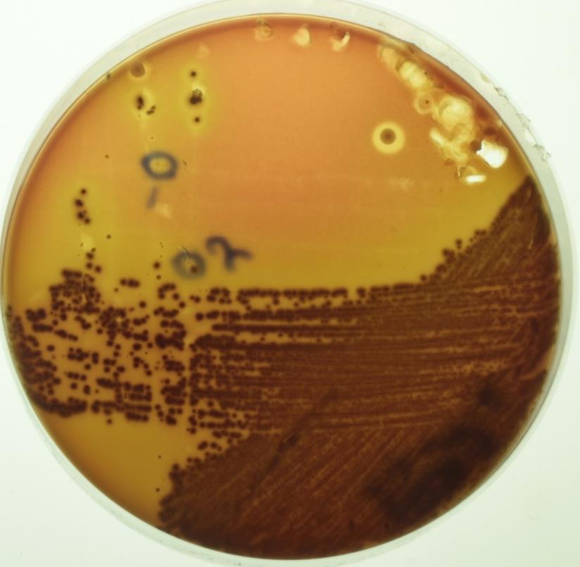
Приложение 1
Фотография смешанных культур факультативных анаэробов: посев материала от пациента № 20 (143) (произведен рассев методом «истощающего штриха» (по Дригальски).
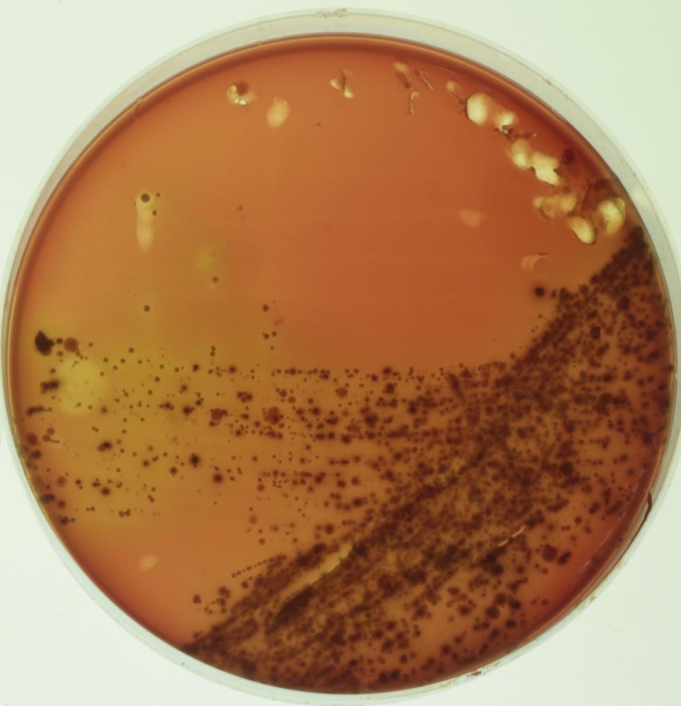
Приложение 2
Фотография смешанных культур облигатных анаэробов: посев материала от пациента № 20 (143) (произведен рассев методом «истощающего штриха» (по Дригальски).

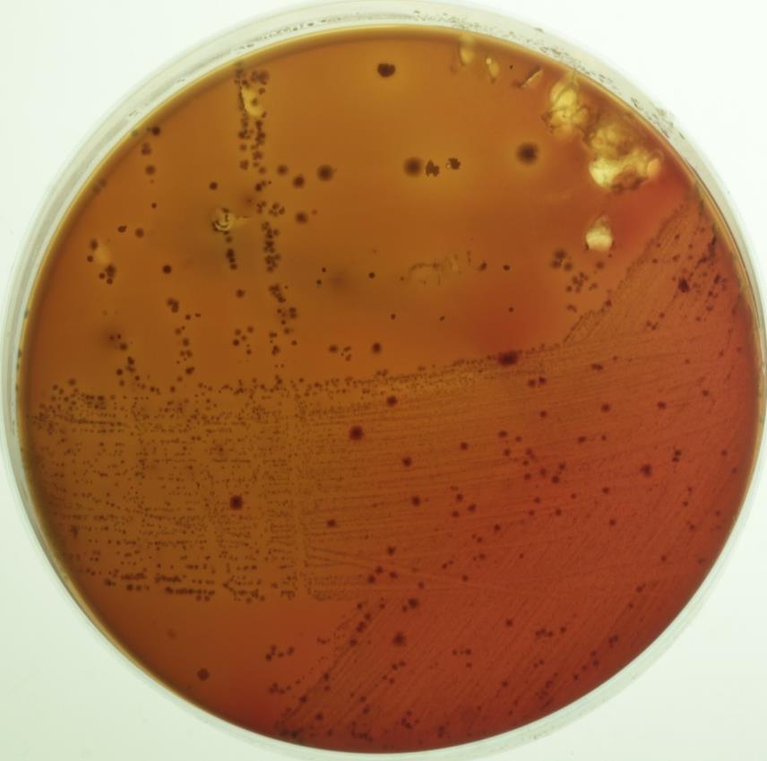
Приложение 3
Фотография смешанных культур факультативных анаэробов: посев материала от пациента № 18 (141) (произведен рассев методом «истощающего штриха» (по Дригальски).
Приложение 4
Фотография смешанных культур облигатных анаэробов: посев материала от пациента № 18 (141) (произведен рассев методом «истощающего штриха» (по Дригальски).
Приложение 5
Материалы VII Научно-практической конференции студенческого научного общества Факультета Стоматологии и медицинских технологий СПбГУ: копия стр.1
Приложение 6
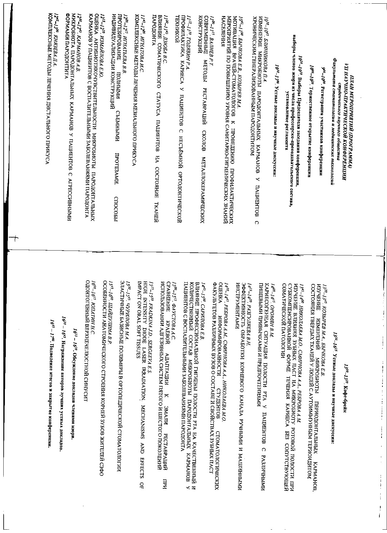
Материалы VII Научно-практической конференции студенческого научного общества Факультета Стоматологии и медицинских технологий СПбГУ: копия стр. 4-5 (программа конференции)
Приложение 7
Материалы VII Научно-практической конференции студенческого научного общества Факультета Стоматологии и медицинских технологий СПбГУ: копия стр. 18-19 (Текст тезисов – начало)
Приложение 8
Материалы VII Научно-практической конференции студенческого научного общества Факультета Стоматологии и медицинских технологий СПбГУ: копия стр. 20-21 (Текст тезисов – продолжение)

|
Из за большого объема этот материал размещен на нескольких страницах:
1 2 3 4 5 6 7 8 9 10 11 |
