

Партнерка на США и Канаду по недвижимости, выплаты в крипто
- 30% recurring commission
- Выплаты в USDT
- Вывод каждую неделю
- Комиссия до 5 лет за каждого referral
The equipment:
1. UHF apparatus.
2. Indicator for investigation of the electrical field.
3. Solutions of electrolyte and a dielectric.
4. Thermometers.
5. Neon lamp.
Electromagnetic Wave
Electromagnetic wave is a diametrical wave, in which a change of electric field tension (E) and magnetic field tension (H) happens in mutual perpendicular planes perpendicularly to the direction of wave distribution (see a fig 1.).
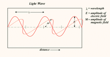
Fig.1. Electromagnetic wave
It was discovered by Maxwell that any alternating electric field creates a vertical magnetic field and vice versa. The mutual creation of the electric and magnetic fields results in the concept of the electromagnetic wave – the circulation of the electromagnetic field in space.
It is known that different electromagnetic wave have the common nature. The electromagnetic oscillations are divided into several groups in dependence of their frequency, that are used with different curative methods:
Low frequency – up to 20 kHz
Sound frequency – 20 Hz – 20 kHz
Ultra-sound frequency – 20 – 200 kHz
High frequency – 0.2 – 30 MHz
Ultra-high frequency – 30 – 300 MHz
Super-high frequency – more 300 MHz
Alternating electric field influence may cause appearance of displacement current and conducting current.
Let's consider the capacity. In the conductor there is the usual current being caused by the charges variation. We can assume that in the capacity this current is replaced by the displacement current, so
![]() ,
,
where ![]()
![]() ,
,
where ![]() – intensity of the electric field,
– intensity of the electric field, ![]() – area of the condenser plane.
– area of the condenser plane.
Magnetic field also influence may change the flowing of physical and biological processes in its tissues. This action is found by Lorenz forces that change the direction of electric charges travel in the inhomogeneous field. Man’s body being placed in the law frequency electromagnetic field (less 100 kHz) has conductor properties and under the influence of electromagnetic field in man’s tissues the conducting currents appear. At the frequencies more than 20 MHz the displacement current prevails.
The law frequency current and electromagnetic fields are used in medicine for electro diagnostic and electro stimulation. At the frequency more then 500 kHz the main action of electromagnetic field is the heating of the tissues. It is used in the physiotherapy and causes a reinforcement of blood flow that connected with arteries and capillaries widening, and decreases blood viscosity as well reinforces filtration and diffusion through biological membranes coming to the removal of proteins from the cytoplasm. Also heating may influence the intermolecular processes causing the specific action in the tissues. In the surgery the high frequency are used to cut and weld tissues.
Electro Conductive Properties of Biological Tissues
In substance under the influence of electromagnetic fields the displacements of electric charges happen. There are free and bounded charges. In conductors free charges prevail. Bounded charges prevail in dielectrics. At the influence of electromagnetic field free charges begin to travel in the conductor and space separation of opposite charges happens or it possibly to say there is the volume polarization of medium. The separated charges shield an outer electric field completely that’s why the electric field is absent inside of the conductor. In dielectrics under the influence of electromagnetic field the displacement of opposite electric charges relatively each other within the limits of an atom or molecule happens. The electric field that created by this charge distribution weakens an outer field just partly.
The main influence is created by the electrical component of electromagnetic field. It causes alternating polarization of dielectric and at the present of electro-conductivity-alternating electric current. The magnetic component creates indirect due to the voltage electric field causing vertical currents.
The action of the alternating current on the organism depends on its frequency. The first method used in medicine practice at the beginning of the 20th century was d’arsonvalization.
D’Arsonvalization Electromagnetic oscillations at the frequency ~ 500 kHz are used at the d’arsonvalization. The curative effect is due to the weak irritation of the skin receptors by electric charges.
Diathermy is a method at which alternating current (current frequency – 0.5-2.0 MHz) is passed through human’s tissues. Biological effect – secrete of heat is found by electrical component of the electromagnetic field. The heat amount that is released in time unit per volume unit of tissue, ΔQ, is found so: ΔQ ~ j2/λ, where j – current density, λ – specific electro conductivity of tissue. Heating and irritation is due to the dipole displacement in dielectric.
During many years diathermy was the main method of high frequency therapy at which heating of inner organs and tissues possibly.
There is a surgery diathermy. This method allows destroying biological tissues at the passing of high frequency electric current.
Inductothermy is a curative method by heating. At this method high frequency magnetic field is used (10-15 MHz). Alternating magnetic field is used for heating of current conductive tissues, i. e. in liquids – blood, lymph. The heat amount that is released in time unit per volume unit of tissue, ΔQ, is found so: ΔQ ~ λνB2, where λ – specific electro conductivity, ν – frequency of oscillations, B – induction of magnetic field.
Ultra High Frequency (UHF) therapy is a curative heating at which high frequency electric field is used (20-50 MHz). This kind of electric field causes a conducting current in the conductive medium and a displacement current in the dielectric. At the UHF therapy the heat is secreted in tissues that have properties of electrolytes as well in tissues with dielectric properties.
The heating of tissues – electrolytes (conductors) in the UHF field happens due to the conducting currents.
It’s necessary to calculate the heat amount releasing in different tissues to appreciate the efficacy of this method. The power of electric current may be represented by: ![]() , where
, where ![]() - specific resistivety,
- specific resistivety, ![]() - distance between condenser planes,
- distance between condenser planes, ![]() - area of condenser plane. Transforming this formula we can get the heat amount that is releases in time unit per volume unit of tissue, ΔQ is found so:
- area of condenser plane. Transforming this formula we can get the heat amount that is releases in time unit per volume unit of tissue, ΔQ is found so:
![]()
where ![]() - electric field tension.
- electric field tension.
In dielectrics the heat is secreted under the influence of alternating electric current due to the change of the orientation of molecule dipoles. The heat amount that is released in time unit per volume unit of tissue,
![]()
where ![]() - tangent of angle of dielectrical losses that characterizes the dipole inertia and depends on dielectric nature (see fig. 2),
- tangent of angle of dielectrical losses that characterizes the dipole inertia and depends on dielectric nature (see fig. 2), ![]() - dielectrical permeability.
- dielectrical permeability.
 |
At the UHF therapy the heat amount secreted in dielectrical tissues is more than electro conductive ones.
In common case the summary heat effect at the influence of UHF field is:
![]()
Order of Carrying Out of the Laboratory Work
1. Switch on UHF apparatus.
2. Find a resonance using a neon lamp.
3. To place ditches with solutions between electrodes.
4. After every 5 minutes to register value of temperature for each solution.
5. Put all data in table.
6. Use the obtained data for making the diagram of the dependence of the temperature from time.
№ | τ, min | Temperature, t0C | ||
Physiological solution | Distilled water | Oil | ||
1. | ||||
2. | ||||
3. | ||||
4. | ||||
5. | ||||
6. | ||||
7. |
8. Write you conclusion.
Laboratory Work №9
The Physical Basis of Electrocardiography. Method of Taken Electrocardiogram
The aim of this work:
1. To study the physical basis of electrocardiography
2. To take the electrocardiogram using electrocardiograph
3. To process the cardiogram
4 Calculating the pulse using electrocardiogram
The equipment:
1. Electrocardiograph
2. Electrodes
3. Physical solution
4. Linings under electrodes
Electric Dipole
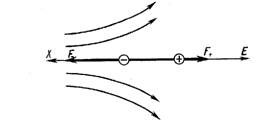
Consider two charges +q at A and –q at B are separated by a small distance r (see the scheme):
![]()
![]()
![]() +q - q
+q - q
P
This configuration is called an electric dipole. Thus two dissimilar charges separated by a small distance constitute an electric dipole. The magnitude of the electric dipole is given by the dipole moment P and is a product of one of the charges and a distance between them. Sometimes this distance is called a brachium of dipole (L).
P = q L
|
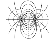
The direction of P is from –q to +q and unit is Coulomb meter (Cm). There is an electric field around dipole in a dielectric (see the figure 1). In the figure: the solid lines are the lines of electrical intensity (force of electrical field)); the dashed lines represent equipotent surfaces.
The Important Properties of Lines of Forces are Listed Below:
· Lines of force start from positive charge and terminate at negative charge.
· Lines of force never intersect each other.
· The tangent to line of force at any point gives the direction of the electric field E at that point.
· The number of lines of force per unit area at right angles to the lines is proportional to the magnitude of E. If E is large, lines are closer together. If E is small, the lines of force are far apart.
In an electric field we think of points to possess electric potential analogous to gravitational potential. In the field around a positive charge, for example, a positive charge moves from points nearer the charge to points farther away. Thus, points around the charges are said to have different electric knowing the electric potential at a point in an electric field, the intensity at that point can be obtained easily.
 In general, the electrical potential at a point is defined as amount of work done in moving a unit positive charge from unity to that point against electrical forces. In a plane of the figure the equipotent lines figure surfaces. The central surface represents a plane transiting perpendicularly to a brachium of a dipole through its middle. All its points have zero potential.
|
Dipole in an Electric Field
The dipole is not only a source of an electric field, but also itself can interact with an exterior electric field.
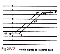
а) The dipole in a uniform external electric field.
According to the figure 2, the dipole is placed in a uniform electric field E. Let the dipole makes an angle θ with the field. The force at +q is F=qE and the force at –q is F=-qE. Hence two forces are equal and opposite each other, therefore the net force is zero. But there is a net torque (moment of force) about the center of a brachium of dipole:
М = qELsinθ
Or in the vector shape:
|
![]()
Thus an electric dipole placed in an external electric field E experiences a torque tending to align the dipole according to the field.
b) Dipole in a non-uniform electric field.
The rotary activity has a place in a non-uniform electric. However, the forces F+ and F - are not equal to each other (see fig.3).
There is a force retracting a dipole into area of a stronger electric field. The quantitative value of this force depends on a gradient of intensity along horizontal axis. If the dipole with the moment Р is oriented along axis X, the net force is determined by the formula:
|
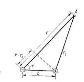
F = P dE/dх.
Electric Field of a Dipole
Dipole is a source of an electric field. The quantitative value of this electric field depends on the dipole moment Р, inductivity of medium ε and some geometrical parameters. Let a dipole be placed in a dielectric infinite medium. A point A is remote from center of dipole on distance r >> L. Let's designate as α angle between a vector Р and direction to this point A. Then potential of the dipole electric field in the point A, is determined by the following formula:
φ = P cosα /(4πεε0r2)
Dipole in an Equilateral Triangle
If a dipole is located in the centre of an equilateral triangle, the beginning of the dipole moment vector will be equidistant for all of its vertexes (see the figure4).
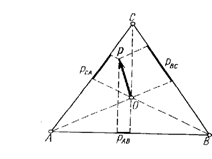
|
It is possible to show, that potential difference (voltage) between two any vertexes is proportional to projections of the dipole moment on the matching side of the triangle:
РАС : РАВ : РСВ = UАС : UАВ : UСВ
Therefore we can judge about the dipole moment vector and its locating inside a triangle if the projections of the dipole moment on the matching side are known. This idea is used in the method of electrocardiography.
The Current Dipole
The electric dipole can be kept a long time in vacuum or in an ideal dielectric. However there is a motion of free charges in real conductive medium. So the dipole will be surrounded by free charges and as a result the dipole is screened:

 + + - -
+ - + - + -
+ + - -
|
There is electrical current in electrical circuit. Such bipolar system is termed the dipole electrical generator or current dipole. The dipole moment of the current dipole is determined by the formula:
Рc = I L,
here L – is the distance between electrodes of the current dipole, I –is an electric current. The electrical potential of the current dipole in point A equals:
φ = Рc cos α / (4 πg r2)
here g – is a characteristics of conductive properties of medium and is termed specific electric conductivity ( g= 1/ρ , ρ - is specific electrical resistance).
The Physics Basis of Electrocardiography
The living tissue is a source of electrical potentials. Electrography is a registration of biological potentials of tissues and organs. There are following diagnostic methods.
ECG - electrocardiography – the registration of biological potentials of cardiac muscle.
ERG - electroretinography - registration of biological potentials of retina in an eye.
EEG - electroencephalography - registration of the electrical potentials of brain.
EMG - electromyography - registration of electrical potentials of muscles.
There are different biological potentials in the table below.
Kind of biological potential | Interval of frequencies, Hz | Amplitude, μV | |
max | min | ||
Electrocardiography | 0, | ||
Electromyography | 1 | 30 - 40 | |
Electroencephalography | 5 - 10 |
Genesis of Electrocardiograms. Eintchoven’s Leads
|
In 1856 Kalliker and Muller detected, that there was potential difference at exaltation of an isolated frog heart. In 1887 an electrocardiogram of a person was registered for the first time. The English scientist Waller with special electrometer made it. The wide
application of electrocardiography began
later, when Eintchoven offered a string galvanometer in 1903. A person’s heart is a potent muscle. The current flows on the surface of a body if this muscle is exited. The potential difference is registrated as electrocardiogram.
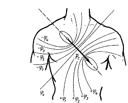
The dipole representation of heart underlies «The theory of Eintchoven’s leads». We consider the heart as a current dipole with the dipole moment Рh. This vector constantly is rotated and changes the position and point of the appendix during a cardiac cycle (see the figure 6).
According to Eintchoven’s theory, the heart is allocated in the centre of an equilateral triangle. The potential differences between triangle vertexes are proportional to the dipole moment projections on the sides of this triangle. These potential differences are named "Eintchoven’s standard leads " in physiology and medicine.
The Eintchoven’s Theory Postulates
1. The heart current dipole is taking place in a center of a triangle. Projections of the dipole moment of the heart are leads of Eintchoven.
2. Electric field of the heart is similar to a field of a current dipole at the big distance from the heart; the dipole moment – is an integrated electric vector of the heart (the sum electric vector of excited cells).
3. The whole organism – is a homogeneous conducting medium (with constant specific resistance).
4.  The direction of heart – current dipole varies during a cardiac cycle, but its point of the appendix doesn’t vary.
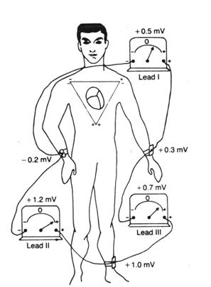
We can see conventional arrangement of electrodes for recording the standard leads in fig.7.
|
of the triangle:
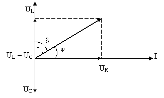
Ul : Ull : Ulll = Рhl : Рhll : Рhlll
The time dependence of a potential difference in leads is recorded as electrocardiogram.
The values of the peak dipole moment of heart in comparison to mass of heart and body are given in the following table.
Object | Mass of the heart, g | body mass, kg | Рh, mА. сm |
frog | 0,16 | 0,036 | 0,005 |
rat | 1,10 | 0,277 | 0,107 |
dog | 108 | 14,2 | 1,63 |
man | 300 | 71,5 | 2,32 |
horse | 3060 | 419 | 13,0 |
The theoretical analysis of electrocardiogram is a complex task. The data of a typical ECG usually supplements a clinical pattern of a disease.

|
The view of a normal electrocardiogram of a person is well known. The electrocardiogram represents the time dependence of potential difference in leads. We can see a potential difference along vertical axis. The horizontal axis is the axis of time. Both electrocardiogram (a) and harmonic spectrum (b) are represented in the figure 8.
The harmonic spectrum is recorded at pulse frequency 66 shocks per minute (period of the heart cycle is 0,9 s).
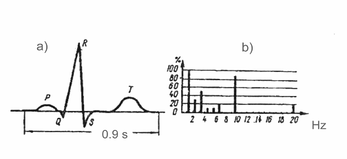
The shape of electrocardiogram has different view in different leads. It is possible to judge about changing of the heart dipole moment Рh by comparing all curves in leads. The ECG is a source of important information. However, the description of electrocardiogram requires good skills in the doctor. The significant simplifying of "reading" of electrocardiogram has become possible with a computer.
Order of Carrying Out of the Laboratory Work
1. Prepare the patient for taken an electrocardiogram.
2. Moisten linings with a physiological solution and fix them to the left hand, right hand, left leg and right leg using electrodes.
3. Connect all electrodes.
4. Switch on electrocardiograph.
5. Push the bottoms on the electrocardiograph and start taken the electrocardiogram.
6. Measure the height of each waves of electrocardiogram.
7. Calculate the potential difference using formula: U = h/S.
8. Put all data in table 1.
Table 1.
№ | Designation | S, mm/mV | H, mm | U, mV |
P Q R S T |
9. Measure a distance l between waves of cardiogram. Calculate a period T, using formula: T = l /υ, where υ – speed of tape a movement.
10. Put all data in table 2.
Table 2.
№ | Designation | υ, mm/s | l, mm | T, s |
R – R P – Q QRS S – T Q – T |
11. Measure pulse rate. Calculate pulse rate using an pared these data.
12. Write your conclusions.
Used Literature
1. Медицинская и биологическая физика. Москва, 1996 г.
2. . Курс физики для медвузов, 1978 г.
3. Практикум по МБФ под ред. проф. Ставрополь, 20стр.
4. Douglas C. Giancoli. Physics. Principles with applications. Fifth edition. Upper Saddle River, New Jersey 07458, 970 p.
5. Dictionary of physics. The penguin. Edited by Valery IllingWorth.
6. Hugh D. Young, Roger A. Freedman. University Physics 9th Edition. Addison Wesley Longman Inc. 1484 p.
7. Марков теории вероятностей и математической статистики. Ставрополь, 20с.
8. Dictionary of mathematics. The Penguin. Edited by John Daintish and R. D. Nelson.
 |

|
Из за большого объема этот материал размещен на нескольких страницах:
1 2 3 4 5 6 7 8 |
